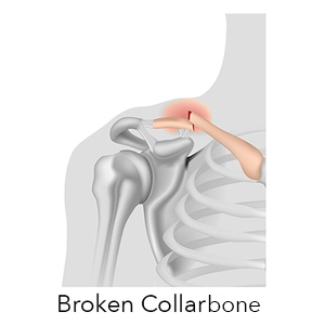

During these visits, he or will take x-rays to make sure the bone is healing in a good position. More strenuous exercises will be started gradually once the fracture is completely healed.įollow-up care. You will need to see your doctor regularly until your fracture heals. These exercises will help prevent stiffness and weakness. Once the bone begins to heal, your pain will decrease and your doctor may start gentle shoulder exercises. Often, patients will begin doing exercises for elbow motion immediately after the injury.After a clavicle fracture, it is common to lose some shoulder and arm strength. Physical therapy. Although there will be some pain, it is important to maintain arm motion to prevent stiffness.Medication. Pain medication, including acetaminophen, can help relieve pain as the fracture heals.Arm support. A simple arm sling is usually used for comfort immediately after the break and to keep your arm and shoulder in position while the injury heals.Most broken collarbones can heal without surgery. If the broken ends of the bones have not significantly shifted out of place, you may not need surgery. Periprosthetic Fracture After Total Hip Replacement.Periprosthetic Fracture After Total Knee Replacement.Congenital Talipes Equinovarus Varus (CTEV) – Clubfoot.Developmental Dislocation (Dysplasia) of the Hip (DDH).Spine Surgical or Non-Surgical Treatment.Medial Patellofemoral Ligament (MPFL) Tear Reconstruction.Posterior Cruciate Ligament (PCL) Reconstruction.Medial Collateral Ligament (MCL) Tear Reconstruction.Anterior Cruciate Ligament (ACL) Reconstruction.Fractures of the Thoracic and Lumbar Spine.Joint Preservation Surgical or Non-Surgical.
#Signs of a broken collarbone skin#
However, the decision to treat a displaced type II fracture operatively in the acute setting may be made if the displacement is so severe that it is at risk of compromising skin integrity.Clavical Fracture (Broken Collarbone) – Dr Mukhi's Raj Hospital Home_MAC 26, 27 Therefore, even displaced fractures can be treated nonoperatively in the same way as types I and III fractures. 1, 14 Despite this, outcome studies have demonstrated similar results in terms of function, strength, and pain in fractures united operatively, those united nonoperatively, and those that go on to nonunion. Because of this displacement, there tends to be a high rate of nonunion. The lateral fracture fragment is held in place by the coracoclavicular ligaments, but pulled downward and medially by the weight of the arm and by the pectoralis and latissimus dorsi muscles, while the medial fragment is pulled superiorly by the trapezius and sternocleidomastoid muscles. Much of the controversy in the literature in treating distal clavicle fractures centers around how best to treat type II fractures, which have a tendency to displace. Most distal fractures can also be treated nonoperatively however, certain factors must be considered in children. Distal fractures are classified based on the relationship to the coracoclavicular ligaments, which determines the likelihood of displacement. Surgery is an option in fractures that have high potential for nonunion (e.g., displaced or communited fractures, fractures with more than 15 to 20 mm clavicle shortening). If a child has no history of trauma, then malignancy, rickets, and physical abuse should be considered.

A prominent callus is common in children, and parents may require reassurance. Most clavicle fractures occur in the midshaft and can be treated nonoperatively. Diagnosis can often be made by the history and physical examination, although appropriate radiography should be used to confirm the diagnosis and guide treatment options. The most common mechanism of injury is a forceful fall with the arm at the side, which commonly occurs during contact sports. Its superficial location, its thin midshaft, and the forces transmitted across it make the clavicle a common site for injury. Clavicle fractures are most common in children and young adults, typically occurring in persons younger than 25 years.


 0 kommentar(er)
